Internal Anatomy
The Water Vascular System
A key feature of echinoderms is a coelomic water vascular system (WVS - Figure 10) composed of a series of complex fluid-filled canals that play a role in respiration, feeding, circulation, locomotion and internal transport (Ruppert et al 2004). As mentioned previously tube feet are part of the external portion of the water vascular system with its roles in feeding, locomotion and respiration; cells within the WVS also collect waste products and transport them to the tube feet where the waste-filled tube feet are pinched off and expelled (Ruppert et al 2004). The inside of the WVS is lined with ciliated (hair-like cells) walls that beat to create a current and along with other cells help to move fluid around the system (Ruppert et al 2004).
The crinoid WVS varies slightly from other echinoderm WVS with two significant differences: stone canals and lack of madreporites (Ruppert et al 2004). The ring canal found within the pentaramous crown gives rise to numerous stone canals, calcareous lined ducts, in between the arms that open into the body cavity (perivisceral coelom) (Ruppert et al 2004). Instead of madreporites, the oral surface of the crinoidis covered by numerous ciliated or tegmental pores that lead, via short integumental (enclosed) canals into the perivisceral coelom (body cavity) (Ruppert et al 2004). Therefore seawater enters the tegmental pores and travels into the perivisceral coelom where it is then pumped into the WVS by the stone canals (Ruppert et al 2004).
Some other key features of a crinoids WVS is the radial canal that comes off the ring canal and diverges into each of the arms, where it then sends a lateral canal into each pinnule which branches into each trio of tube feet (Ruppert et al 2004). The muscle fibers in the radial canal are also contracted by the tube feet to create hydraulic pressure due to the absence of ampullae (Ruppert et al 2004).

Figure 10- An adapted version of the Water Vascular System from Ruppert et al (2004)
Mutable Connective Tissue
Another key feature of echinoderms is their ability to reversibly vary their rigidity using the mutable connective tissues, dermis and general connective tissue, joining the ossicles together (Ruppert et al 2004). The dermis contains numerous cell types, including muscle and nerves cells, but it’s the extracellular matrix within the dermis that allows for the changes in stiffness with the nerves cell terminating within it (Ruppert et al 2004). The extracellular matrix is mainly made up of collagen and elastin, which allows for elasticity, flexibility and strength all important qualities in varying extremes of rigidity (Rupert et al 2004). The nerves present within the extracellular matrix either harden or soften the matrix, with the levels of calcium playing a major role (Ruppert et al 2004). The increase of calcium stiffens the matrix while a decrease softens it, giving rise to the theory that calcium may form connections between the macromolecules within the matrix (Ruppert et al 2004).
Body Wall
The body of a crinoid is capsulated by a cuticle and a non-ciliated epidermis which covers the skeletal ossicle embedded connective tissues of the body below (Ruppert et al 2004). The jointed appearances of the stalk, cirri, arms and pinnules is due to their interiors being packed almost entirely by series of vertebra-like ossicles to make it nearly solid (Ruppert et al 2004). The arm ossicles (brachials) articulate allowing for limited movement while other ossicles within the stalk, cirri and arms are bound by ligaments,collagen fibers, that penetrate into the skeletal material and allow for rigidity changes as seen above (Ruppert et al 2004).
Musculature
Crinoids contain a number of muscular cells with the most distinct muscles occurring mainly in the arms and pinnules while individual muscle fibers are spread throughout the connective tissues of the body and epitheliomuscular cells, cells that contract like muscle, line various parts of the coelomic cavities (Ruppert et al 2004).Between the oral edges of the arm and pinnule ossicles there are obliquely striated flexor muscles that help curl the arms and pinnules while arm (interbrachial) ligaments are responsible for the straightening or extension of these appendages via elastic recoil (Ruppert et al 2004).
Digestive System
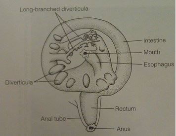
The digestive system of a crinoid is confined to the calyx cup or central disc, with the mouth opening into a short esophagus which leads into the intestines ( Ruppert et al 2004 - Figure 11). The intestines descend and turn around along the inner side of the calyx wall where it then heads upwards into a short rectum which opens into the anus at the top of the anal cone ( Ruppert et al 2004 - Figure 11). The waste products released from the anus are in the form of large, compact, mucus cemented pellets that fall off the anal cone onto the surface of the disc where it then drops off the body ( Ruppert et al 2004). It is believed that both extracellular and intracellular digestion occurs within the intestines but further research is needed to confirm this.
  Figure 11- A diagram of the digestive system of Crinoids modified from Ruppert et al (2004).
 |